CIRCULAR DICHROISM SPECTROSCOPY (CD)
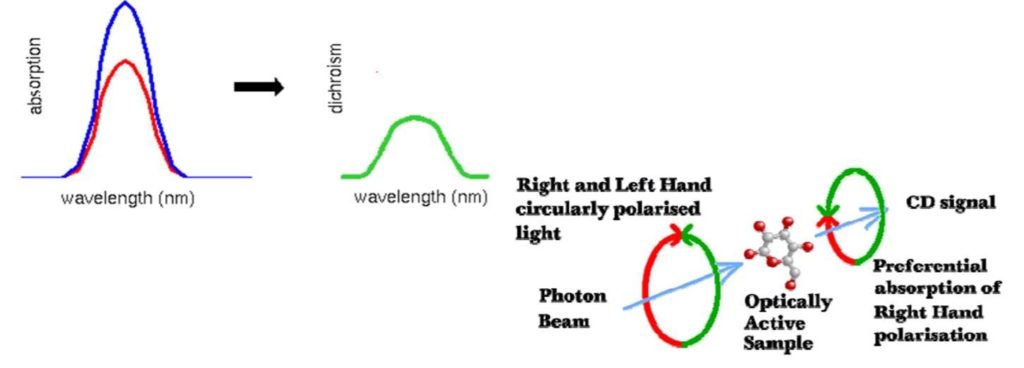
Circular Dichroism (CD) is a type of absorption spectroscopy that can provide information on the structures of many types of biological macromolecules

It measures the difference between the absorption of left and right handed circularly-polarized light by proteins. CD is used for;
- Protein structure determination.
- Induced structural changes, i.e. pH, heat & solvent.
- Protein folding/unfolding.
- Ligand binding
- Structural aspects of nucleic acids, polysaccharides, peptides, hormones & other small molecules.
HIGHER ORDER STRUCTURE ANALYSIS
SECONDARY STRUCTURE ANALYSIS
TERTIARY STRUCTURE ANALYSIS
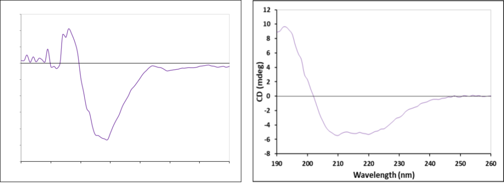
In the “Far-UV” spectral region (190-250 nm) peptide bonds produce CD signals that are sensitive to the overall secondary structure of the protein. By analyzing the CD spectra of Far-UV region, a defined assessment of the components of the protein secondary structure can be made.
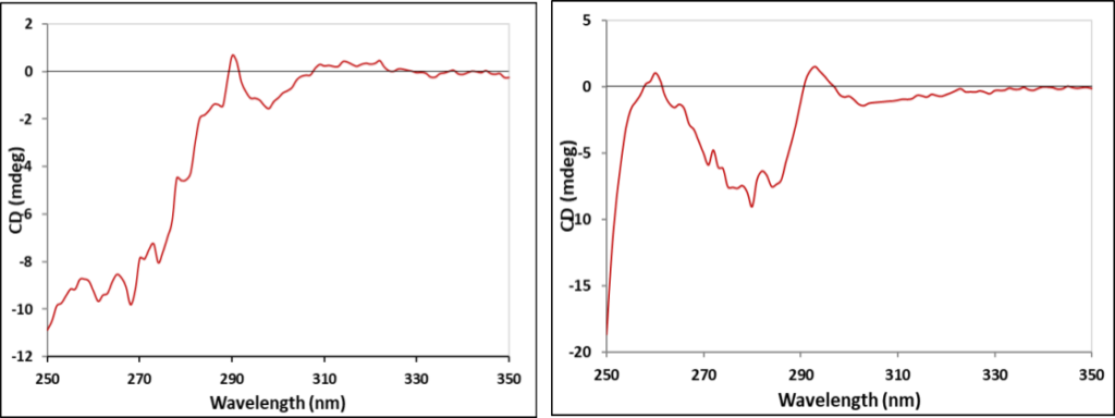
In the “near-UV” spectral region (250-350 nm) aromatic amino acids and disulfide bonds produce CD signals that are sensitive to the overall tertiary structure of the protein. By analyzing the CD spectra of near-UV region,
a good assessment of the tertiary structure of the protein sample can be made.

HIGHER ORDER STRUCTURE ANALYSIS
SECONDARY STRUCTURE ANALYSIS
| -ve band (nm) | +ve band (nm) | |
| α- helix | 222,208 | 192 |
| β- sheet | 216 | 195 |
| β- turn | 220-230 weak
180-190 strong |
205 |
| Random Coil | 200 |
TERTIARY STRUCTURE ANALYSIS
| band (nm) | |
| Phenyl alanine | 250-270 |
| Tyrosine | 270-290 |
| Tryptophan | 280-300 |
| Disulphide bonds | Broad signal throughout Near UV region |

SECONDARY STRUCTURE ANALYSIS
FAR- UV-CD SPECTRA OF Bevacizumab
FAR- UV-CD SPECTRA OF CRM 197

TERTIARY STRUCTURE ANALYSIS
FAR- UV-CD SPECTRA OF Bevacizumab
FAR- UV-CD SPECTRA OF CRM 197

CD ANALYSIS AT CBA
| Secondary structure analysis by Far UV-CD spectroscopy
|
Tertiary Structure Analysis of Protein using Near-UV CD Spectroscopy | |
| Sample requirement | Amount- 0.3-1 ml (0.1-1 mg/ml)
Formulation buffer- 5 ml |
Amount- 1-2 ml (1-2 mg/ml)
Formulation buffer- 5 ml |
| Deliverables | CD Spectra, Secondary structure analysis (relative amount of α-helix, β-sheet and random coil) and MRE (Θ in grad cm² dmol-1) | CD Spectra, MRE (Θ in grad cm² dmol-1) |
| Information required from Client | • Molecular weight
• Number of residues • Concentration • Buffer composition (in case of formulation) This information is essential for the calculation of the optical constants and secondary structural analysis. Note: If the buffer contains substances with high absorbance in the measurement range (e.g. Glycine, Citrate, Benzyl alcohol etc.), this may lead to an overload of the detector and limited measurement range. |
|
SERVICES OFFERED BY CBA
- Protein Intact Mass Analysis using HRMS
- Peptide Mapping using HRMS
- Post Translational Modifications Analysis using HRMS
- Host Cell Protein Impurity Identification using HRMS
- Host Cell Protein Impurity Profiling using SDS-PAGE
- Secondary Structure Analysis using far-UV CD Spectroscopy
- Tertiary Structure Analysis using near-UV CD Spectroscopy
- Tertiary Structure Analysis using Fluorescence Spectroscopy
- Higher Order Structure Analysis using Fluorescence Spectroscopy
- Secondary Structure Analysis using FTIR Spectroscopy
- Higher Order Structure Analysis using DSC
- Aggregate/Multimers Analysis using SEC-MALS
- Protein Protein Interaction Analysis using SPR
- Biosimilarity assessment Project
SERVICES OFFERED BY CBA
Edna Joseph
Head – Analytical Services
Venture Center
100, NCL Innovation Park, Dr. Homi Bhabha Road, Pune – 08
Mobile: +91-7410045651 | Reception: +91-9172232215
Email: cba@venturecenter.co.in
Ms. Rutuja Patil
Assitant Manager – Business Development
Venture Center
100, NCL Innovation Park, Dr. Homi Bhabha Road, Pune – 08
Mobile: +91-8956677542| Reception: +91-9172232215
Email: rutuja.patil@venturecenter.co.in
For more information, please contact:
Website: https://bioanalysis.in/
